Tennis Elbow
Pathology
The main clinical symptoms are pain on resisted movements (particularly resisted third finger extension) and tenderness at the lateral epicondyle, with normal elbow range of motion. The diagnosis is based on the clinical features of the disease. Diagnostic imaging should be considered to rule out other causes of elbow pain or to establish the diagnosis of tennis elbow when in doubt.As with other tendinopathies, the pathology of tennis elbow is complex and not fully understood. Similar to calcifying tendinitis of the shoulder, sudden overload may alter the structure of the tendons at the common extensor origin, leading to a degenerative process. However, calcifications are rare in tennis elbow. The involvement of neurogenic inflammation in Tennis elbow has also been suggested.

Approximately 40% of all tennis players report problems with their elbow, but only a quarter of them consider the symptoms to be disabling and severe. Notably, most patients with tennis elbow do not play tennis. This is due to the fact that many tennis players have a weekly training routine that regularly loads the tendons and keeps them healthy. Rather, the injury usually occurs in people who have been sedentary for years and then overuse a previously underused and atrophied tendon by exercising at the gym, doing gardening, or even just carry heavy luggage. When the injury is caused by playing tennis it is the backhand stroke that leads to excessive loading of the tendons at the common extensor origin.
The population prevalence is approximately 2%, with peak incidence occurring at 40 to 50 years of age.
The initial treatment should be conservative including rest, physiotherapy, and nonsteroidal anti-inflammatory drugs. As in the case of chronic Achilles tendinopathy and chronic plantar fasciopathy, eccentric (lengthening only) exercises have become the mainstay of rehabilitation programs for tennis elbow. In most circumstances, cortisone injections should not be used. This is due to the fact that cortisone leads to very good results in the short term (six weeks) but has been demonstrated to be harmful in the longer term (more than three months). Surgery should be considered when conservative treatment fails.
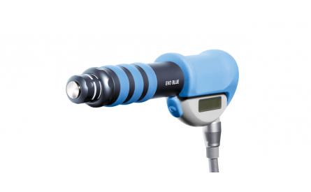
An attractive alternative to surgery is Radial Shock Wave Therapy for tennis elbow treatement.
Side effects of Radial Shock Wave Therapy (RSWT) using the Swiss DolorClast®.
When performed properly, RSWT with the Swiss DolorClast® has only minimal risks. Typical device-related non-serious adverse events are:
- Pain and discomfort during and after treatment (anaesthesia is not necessary)
- Reddening of the skin
- Petechia
- Swelling and numbness of the skin over the treatment area
These device-related non-serious adverse events usually disappear within 36h after the treatment.
Treatment Procedure
Locate the area of pain through palpation and biofeedback.
Mark the area of pain.
Apply coupling gel to transmit shock waves to the tissue.
Deliver Radial or Focused Shock Waves to the area of pain while keeping the applicator firmly in place on the skin.

