Back to top

You are here
01 - Assess & engage

SEE ALL
CHAPTERS

Assess & engage

How to recognise plantar fasciopathy?

The patient’s history, together with the physical examination of the foot and ankle, should provide valuable insights regarding the underlying pathology. Common red flags include:

  • patients present walking with their affected foot in equinus position to avoid applying pressure to the painful heel, or ‘‘walking on a stone’’; 
  • hyperpronation of the affected foot;
  • obesity;  
  • patients demonstrate feelings of discomfort in the proximal plantar fascia with passive ankle/first toe dorsiflexion;
  • patients report pain occurring when taking their first steps in the morning or after periods of rest; 
  • the presence of tenderness on the medial calcaneal tuberosity and along the plantar fascia.
     

Although plantar fasciopathy is the most common source of plantar heel pain, the differential diagnosis of plantar fasciopathy is broad, encompassing arthritic conditions, infections, neuropathies, and mechanical causes. Mechanical etiologies of plantar heel pain include:


Physical examination involves:

  • Palpation of the medial tubercle of the calcaneus and proximal plantar fascial insertion at the anteromedial calcaneus. 
  • Passive ankle dorsiflexion. Stretching of the plantar fascia by passive dorsiflexion results in an increase in pain.
  • The Windlass test involves forced dorsiflexion of the toes at the metatarsophalangeal joints with the ankle stabilized. Reproduced heel pain indicates a positive result. 
     
plantar fasciopathy gif palpation


Imaging

  • Although generally unnecessary, guidelines from the American College of Radiology recommend weight-bearing radiography as an initial test to rule out other causes of heel pain.
  • MRI makes it possible to demonstrate the thickening of the proximal plantar fascia. Increased proximal plantar fascia thickening with augmented signal intensity on T2-weighted and short tau inversion recovery images are suggestive of plantar fasciopathy. 
  • Diagnostic medical sonography has been found to be effective in differentiating the normal plantar fascia from tissue affected by plantar fasciopathy, thus allowing soft tissue pathology of the heel to be ruled out. Ultrasound findings suggestive of plantar fasciopathy involve plantar fascia thickness greater than 4.0 mm and reduced echogenicity of the plantar fascia. 
Image
heel spur
Heel spur

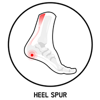
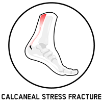
Confirmed by radiographic findings at the pain location. As many as 50% of patients with plantar fasciopathy have heel spurs. A heel spur is usually an incidental finding that is not diagnosed on the basis of the patient’s symptoms [1,3]. 

Image
calcaneal stress fracture
Calcaneal stress fracture

Pain during activity, progressing to pain at rest.
 

Image
nerve entrapment
Nerve entrapment

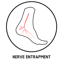
(medial or lateral plantar nerve, nerve to abductor digiti minimi). Characterized by feelings of burning, tingling, or numbness. 
 

Image
heel pad syndrome
Heel pad syndrome

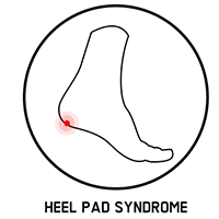
 manifested by deep, bruise-like pain, is typically localized in the middle of the heel. 
 

Image
tarsal tunnel syndrome
Tarsal tunner syndrome

manifested by a sensation of pain, burning and tingling on the sole of the foot. 

Treatment of plantar fasciopathy

The treatment of plantar fasciopathy and the alleviation of pain to increase the patient’s quality of life requires: 

  • suppression of inflammation;
  • stimulation of fascia healing;
  • functional improvement.

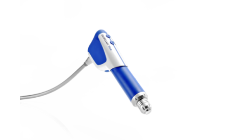
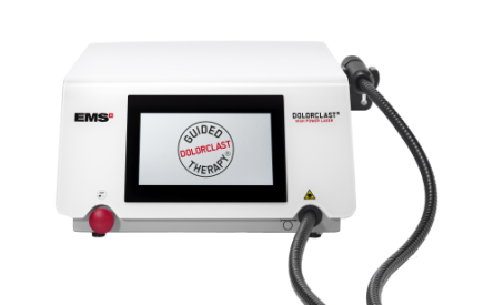
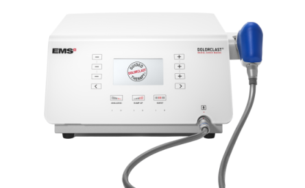
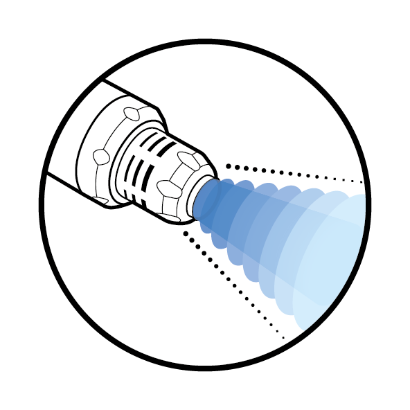
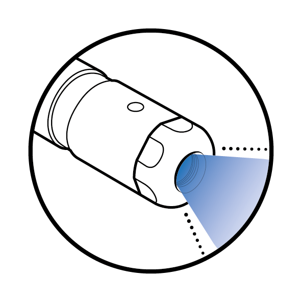
The treatment of plantar fasciopathy can include pharmaceutical and non-pharmaceutical methods such as Guided DolorClast® Therapy. It is recommended to begin the therapy with a non-pharmacological and non-invasive approach, such as shock wave therapy, physical exercise, and patient education, before pharmacological interventions. Opioids and steroids may reduce pain and inflammation, but they do not improve function and are associated with side effects, such as nausea and headache.


What causes pain in plantar fasciopathy?

Pain can have multiple origins, such as mechanical strain, degeneration of tissues, inflammation, and edema. The tendency is often to treat the dominant symptom – in this case, pain – without resolving its cause. With quick fixes such as painkillers, underlying inflammation can be neglected, preventing tissue healing. Although the patient may be satisfied with the recession of pain, this may ultimately lead to greater damage due to the stress that continues to be applied to the injury. Compared to other approaches, radial shock waves effectively reduce pain and inflammation with no side effects.

As many as 20% of plantar fasciopathy patients experience persistent symptoms and unsatisfactory treatment outcomes [5-6]. A recent meta-analysis comparing as many as seven different treatment modalities in plantar fasciopathy patients concluded that RSWT® offered more effective and stable pain relief compared with other interventions [5].


Bibliography
+
[1] League, Alan C. “Current concepts review: plantar fasciitis.” Foot & ankle international vol. 29,3 (2008): 358-66. doi:10.3113/FAI.2008.0358
[2] Ibrahim, Mahmoud I et al. “Chronic plantar fasciitis treated with two sessions of radial extracorporeal shock wave therapy.” Foot & ankle international vol. 31,5 (2010): 391-7. doi:10.3113/FAI.2010.0391
[3] Petraglia, Federica et al. “Plantar fasciitis in athletes: diagnostic and treatment strategies. A systematic review.” Muscles, ligaments and tendons journal vol. 7,1 107-118. 10 May. 2017, doi:10.11138/mltj/2017.7.1.107
[4] Thomas JL, Christensen JC, Kravitz SR, Mendicino RW, Schuberth JM, Vanore JV, Weil LS Sr, Zlotoff HJ, Bouché R, Baker J; American College of Foot and Ankle Surgeons heel pain committee. The diagnosis and treatment of heel pain: a clinical practice guideline-revision 2010. J Foot Ankle Surg. 2010 May-Jun;49(3 Suppl):S1-19. doi: 10.1053/j.jfas.2010.01.001. PMID: 20439021.
[5] Li, Xian et al. “Comparative effectiveness of extracorporeal shock wave, ultrasound, low-level laser therapy, noninvasive interactive neurostimulation, and pulsed radiofrequency treatment for treating plantar fasciitis: A systematic review and network meta-analysis.” Medicine vol. 97,43 (2018): e12819. doi:10.1097/MD.0000000000012819
[6] Goff JD, Crawford R. Diagnosis and treatment of plantar fasciitis. Am Fam Physician. 2011;84(6):676-682.

Looking for a treatment? Find a practice for a treatment near you.

Looking for a treatment? Find a practice for a treatment near you.